
Ultrasound Differential Between Fetal Hydrops and Cystic Hygroma
Fetal hydrops is a condition in fetus that shows fluid
accumulation (edema) in two or more fetal compartments, fluid accumulation can be seen in:
- Abdominal cavity (ascites)
- Pericardium (pericardial effusion)
- Pleural cavity (pleural effusion)
- Generalize edema
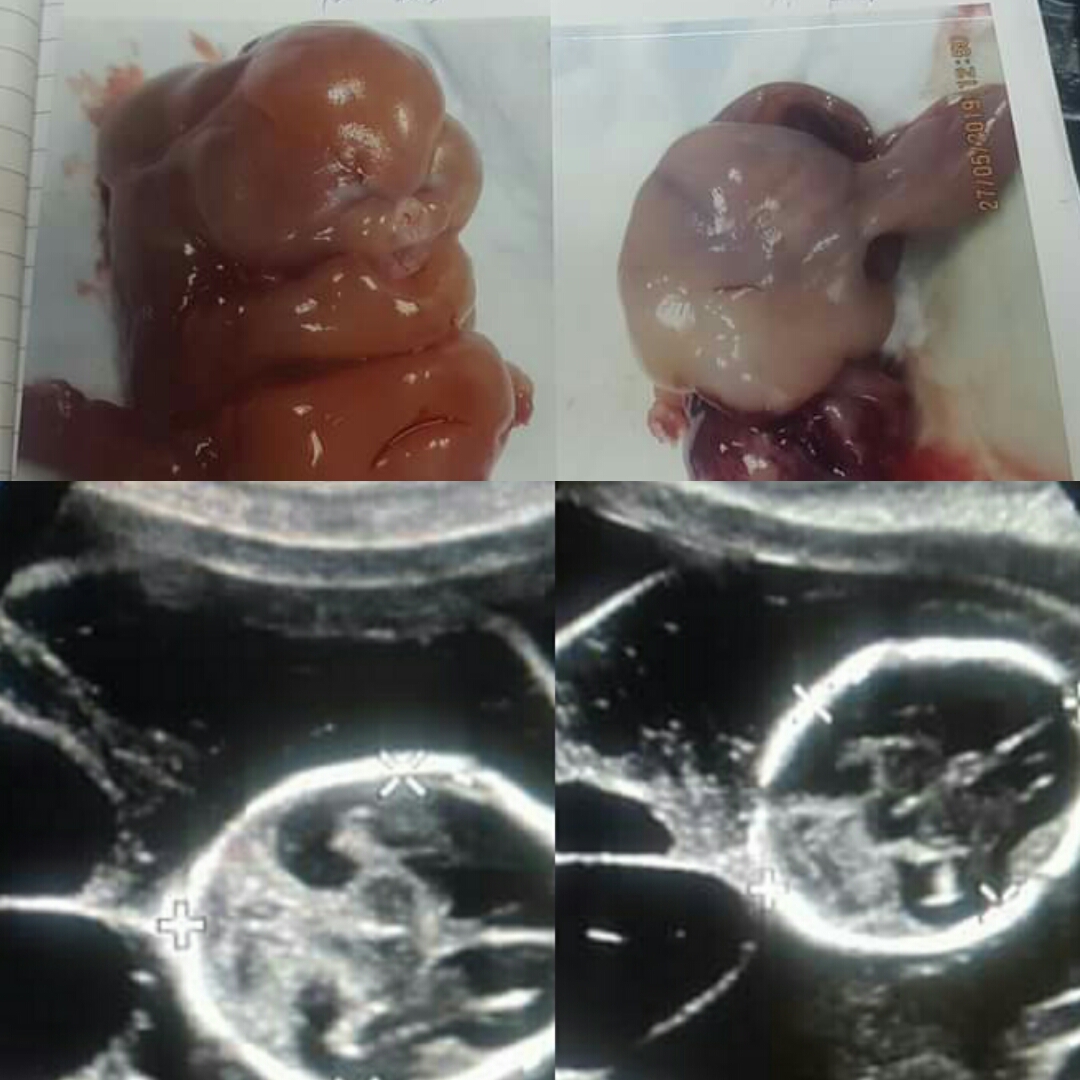 |
| Picture shows aborted edematous fetus |
Fetal hydrops is not a disease but an ultrasound indicator
for other intra-uterine complications. Fetal hydrops can be immune or
non-immune related in fetal anemia, however non-immune hydrops can be
independent of anemia in which case tumor or congenital cystic adenomatoid
malformation cause increase blood flow demand. This over-bearing cardiac output
leads to edema secondary to fetal heart failure.
Immune fetal hydrops is seen in unmanaged Rh disease. This
incident has since reduced with an improved Rh disease management.
Fetal cystic hygroma is a congenital malformation of the
lymphatic system appearing as an infiltrative cystic mass with single or
multiloculated fluid filled cavities and variable density of fluid, soft tissue
and fat combination. They are caused by lymphatic system blockage and usually
around the neck. This usually results to hydrops. Cystic hygroma can
be caused by both genetic and environment factors such as:
- Viral infection
- Drug or alcohol use during pregnancy
- Some chromosomal abnormality

Things to have in mind for differential considerations during antenatal ultrasound includes:
- Cervical teratoma
- Occipital encephalocele
- Cervical meningocele


Comments
Post a Comment